The immune cell profiler
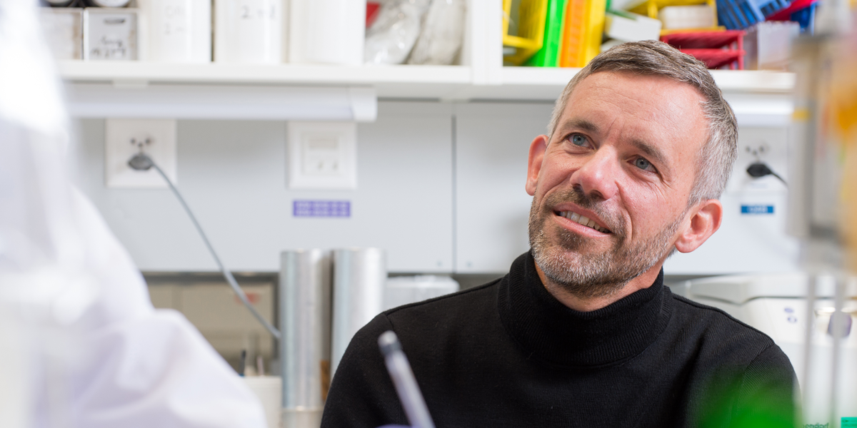
Mikaël Pittet started out as a novice researcher at Ludwig Lausanne in 1998. He has returned as a full Member of the Ludwig Institute and an accomplished immunologist exploring how the various functional states of myeloid cells—frontline soldiers of the innate immune response—influence antitumor immunity and immunotherapy.
When Mikaël Pittet was about 15 years old, his mother, a nurse by profession, took him to Epalinges, a suburb of his hometown of Lausanne. Someone she knew knew someone known to be a renowned scientist and, having noticed her son’s incipient fascination with science, she had asked—and the scientist had graciously arranged—to have young Pittet visit his laboratory. The scientist was immunologist Jean-Charles Cerottini, then director of the Lausanne Branch of the Ludwig Institute for Cancer Research and a pioneer in the study of the immune system’s T cells. Pittet wound up visiting Ludwig Lausanne for a full week. “And so my very first exposure to science, when I was still a kid, was at the Ludwig Institute,” says Pittet. “I was impressed.”
And, apparently, inspired. Not only would Pittet turn to immunology for his graduate studies at the University of Lausanne, but he would also conduct his doctoral research—and his postdoctoral fellowship—at Ludwig Lausanne under the mentorship of none other than Cerottini and the immunologists Pedro Romero and Daniel Speiser before leaving for the U.S. in 2003. “I was fascinated by the vision that Jean-Charles had for how the immune system could be harnessed to fight cancer,” says Pittet. “This was obviously long before it was fashionable.”
In the years since, Pittet has only burrowed deeper into the complexities of tumor immunology, beginning as a professor at Harvard Medical School and, since 2020, at the University of Geneva, holding the ISREC Foundation chair of immuno-oncology. His laboratory, now at the AGORA Cancer Research Cluster in Lausanne, has become an epicenter for the study of myeloid cells—including neutrophils, dendritic cells and macrophages—associated with tumors. Pittet has teased apart unique states assumed by these frontline soldiers of the innate immune response and detailed how they can support tumor growth and survival, interfere with immunotherapies or boost anti-tumor immunity, depending on their functional flavors.
In 2021, Pittet began his third stint at Ludwig Lausanne, this time as a full Member of the Ludwig Institute.
GETTING HOOKED
For a fledgling scientist, few experiences could have been more inspiring than joining the team at Ludwig Lausanne in 1998. For one thing, Pittet got to travel with his mentors to meetings of Ludwig researchers from around the world hosted in New York by the late Lloyd Old, former Ludwig CEO and scientific director.
“Being able to interact with this giant of cancer research was amazing,” recalls Pittet, who was no less dazzled by the intellectual firepower Old convened at the New York meetings. “It was just crazy as a young scientist to meet people whose names I’d seen on papers,” he says. “It gave me the additional motivation to do the best I could to also contribute a little bit to the science. I was hooked.”
And then there was, of course, the work itself. Ludwig Lausanne researchers had adapted and developed a technology, known as tetramer assays, to rapidly detect anti-tumor T cells isolated from patients. For his doctoral research and his subsequent postdoctoral fellowship in Romero’s lab, Pittet used the technology to identify tumor-reactive T cells isolated from melanoma patients and analyze responses to a cancer antigen known as Melan-A. Aside from their intrinsic significance to tumor immunology, these studies entailed the development of methods that have since been widely adopted to assess T cell responses to immunotherapy.
Most significant for Pittet, however, were the larger lessons garnered from his studies. His findings helped establish that anti-tumor T cell responses were indeed occurring naturally in melanoma patients—that they weren’t just artifacts of experimentation. Yet, notably, these killer T cells also seemed to be largely ineffectual within the tumor. Intriguingly, when those same, lethargic T cells were put in a dish and fed certain stimulatory immune factors known as cytokines, they revived their cancer killing function within a day.
“So there was this notion that there would be some reversibility in the T cell suppression, or anergy, as we called it at the time,” says Pittet. “We now know how important this was, considering the capacity to activate or reactivate T cells with immunotherapeutic agents.”
THE IMPORTANCE OF LOOKING
It was this suppression of the anti-tumor response and its reversal that Pittet decided he would explore next. For this, he reasoned, he’d need to pursue both imaging in live animals and mechanistic studies. “I visited labs that were best known for their imaging capabilities and ended up going to the Center for Molecular Imaging Research at Massachusetts General Hospital, which was led by Ralph Weissleder, who became a great mentor,” says Pittet. “I‘ve worked closely with him for 17 exciting years.”
In Boston, Pittet began exploring how killer T cells are suppressed in the tumors of mice. His real-time imaging studies—done with Weissleder, Thorsten Mempel and Ulrich von Andrian at Harvard Medical School and Harald von Boehmer and Khashayarsha Khazaie at the Dana Farber Cancer Institute—showed that immunosuppressive regulatory T cells (Tregs) in tumors monkey-wrench the engagement of the killer T cell’s weaponry, specifically, the release of cytotoxic granules into target cells. Their mechanistic analyses showed the effect to be dependent on a factor known as TGF-β. Reported in 2005 and 2006, these studies were among the first to capture how precisely Tregs repress killer T cell function. Also notable was the discovery that the killer T cells themselves retained their cytotoxic capabilities and that their suppression could be reversed.
THE ORIGINS OF THINGS
Setting up his new laboratory at Massachusetts General Hospital in 2006, Pittet decided he was ready to move on from T cell immunology. “I realized that tumors are complex entities, and while focusing on T cells was important, they represent just one part of an intricate ecosystem,” he explains. Of all the noncancerous cell types in tumors, Pittet chose myeloid cells as a worthwhile focus because they tend to be so numerous in tumors and because, being an immunologist, they felt somewhat closer to him.
But he first a took a detour, turning his attention for a spell to the role of these cells in chronic and acute inflammation in mouse models of myocardial infarction and atherosclerosis. These studies demonstrated that subsets of macrophages change dramatically in inflammatory conditions related to cardiovascular disease. They also identified myeloid cells—like monocytes, and the macrophages derived from them—causally involved in such things as atherosclerosis and the healing of heart muscle. Perhaps the splashiest discovery Pittet’s lab made in this arena was the identification in 2009 of the much-neglected spleen as a vital reservoir of monocytes that heal cardiac muscle after a heart attack.
That fascination with the origins of myeloid cells persisted when Pittet turned once again to cancer research. Macrophages, including those that support tumor growth, derive in part from monocytes in the blood, which in turn are generated by hematopoietic stem cells in the bone marrow. “We wondered, could it be that hematopoietic [blood-forming] stem cells are regulated by cancer?” says Pittet. “Is there long-range communication between cancer and other locations in the body? And that became very important for my lab—seeing cancer as a systemic disease, one that can have an impact far away from its location.”
In 2012, Pittet’s lab showed in a mouse model of lung cancer that the spleen also serves as a site for the production of macrophage and neutrophil precursors, which are then sent to tumors where they promote malignant growth. A year later, he and his colleagues reported that tumors can manipulate a hormonal circuit controlled by the blood pressure-regulating hormone angiotensin-II to remotely drive the production of macrophage and neutrophil precursors. Another study, published in 2017, showed in both mouse models and cancer patients that lung tumors—even before they’ve metastasized to bone—can remotely influence certain marrow cells to drive the production of a subtype of neutrophils that strongly promote cancer growth.
These studies continue today. “They represent one facet, or one half, of the work in my lab—these fundamental studies connect cancer to the immune system in the entire organism, not just in the tumor,” says Pittet.
The other half relies on sophisticated real-time imaging in living animals to figure out how drugs work.
THE THIEVING MACROPHAGE
These pharmacokinetic and pharmacodynamic studies—combined with the analysis of global gene expression in individual cells—have opened a unique window into the influence tumor-associated myeloid cells have on therapy.
“When we give a drug to a patient or a mouse, we still know little about why it sometimes works and sometimes fails,” explains Pittet. “We want to address this black box, to literally illuminate it using fluorescence imaging and microscopy. We want to be able to see the drugs, tumor cells and key immune cells and molecules involved in anti-tumor immunity, and we want to see all this in real time.”
Seeing—not merely deducing—what goes on inside a tumor can make all the difference. Tracking anti-PD-1 antibodies in real time in a mouse model of cancer, for example, Pittet and his colleagues found that the drugs were binding their targets on killer T cells within tumors, presumably disengaging the brakes on their anti-tumor activity. But what they saw next surprised them. When the T cells bearing the antibodies bumped into a tumor-associated macrophage, the latter stole the antibody off the surface of the T cell. “This is not a good thing because the T cell can no longer be fully activated, which prevents the treatment from being fully effective,” says Pittet.
The more such macrophages there were in tumors, the more likely the drug would be stolen away. Pittet and his colleagues reported these findings in 2017, revealing that a receptor normally expressed by macrophages was snaring the antibodies. Further, blockade of that receptor could boost the efficacy of anti-PD-1 immunotherapy in mouse models, suggesting a novel strategy to enhance checkpoint blockade.
A WELCOMING COMMITTEE
Pittet’s lab has also explored the determinants of success for checkpoint blockade. Imaging studies using intravital microscopy revealed that treatment with anti-PD-1 antibodies caused a massive activation within tumors of a population of dendritic cells—which direct and stimulate the T cell attack. Further analysis indicated that this newly identified state of intratumoral dendritic cells is relatively rare but absolutely essential for effective checkpoint blockade.
When killer T cells are activated by anti-PD-1 antibodies, they produce a protein factor known as interferon (IFN)-γ. Mechanistic analysis revealed that IFN-γ prompts this state of dendritic cells to produce an immune factor, interleukin-12, that is sensed by the T cells. “The production of IL-12 inside the tumor tells the T cells that they can go kill their target,” explains Pittet. In addition, these dendritic cells tend to congregate around the blood vessels feeding tumors. “When a T cell arrives in the tumor, the first cells it sees are likely to be these dendritic cells, which are strategically positioned—like a welcoming committee,” says Pittet.
Since publishing these findings in 2018, Pittet’s lab has also reported that the dendritic cells produce a factor that retains T cells in their niche and another, interleukin-15, that is known to promote T cell survival. Since their discovery of these dendritic cells, other labs have independently identified the same cells. What they should be named, however, remains up in the air. “The field is still in its infancy,” says Pittet.
THE NEGLECTED NEUTROPHIL
Similar studies done on the types of neutrophils within tumors, meanwhile, have revealed one state that promotes tumor growth and another that has antitumor activity. “A few years ago, neutrophils were mainly considered a homogeneous population whose role in cancer was not clear,” says Pittet.
Analyzing the gene expression patterns of individual neutrophils—in work done in collaboration with Harvard researcher Allon Klein—his lab found that there are multiple states of neutrophils associated with lung tumors. Further, as they reported in 2019, the various states recur across patients and species, suggesting that interventions to manipulate specific neutrophil states in mice are likely to hold in humans and also to be of general benefit to lung cancer patients.
Dissecting one of these neutrophil states, Pittet and his colleagues identified a population whose presence in the tumor microenvironment is consistently associated with poor patient outcomes. This neutrophil state tends to be very long lived. “We believe these cells could be an important immunotherapeutic target because they have all the characteristics of a tumor-promoting cell, are present in many patients, sometimes in very high frequencies, and are largely ignored from a therapeutic perspective,” says Pittet.
LOOKING AHEAD
As a tumor ecologist of sorts, Pittet finds himself in excellent company at Ludwig Lausanne. Other researchers at the Branch, most notably in the laboratories of Member Johanna Joyce and Associate Member Ping-Chih-Ho, are interested in different aspects of the role played by myeloid cells in cancer.
Pittet is also collaborating with Branch Director George Coukos to examine the promise of a novel approach to cancer therapy, FLASH radiotherapy, being developed at the Lausanne University Hospital (CHUV). The approach employs novel technology to target tumors with extremely high doses of radiotherapy while sparing healthy tissues. He and Coukos will be examining how the immune system might be recruited to destroy tumors during FLASH radiotherapy. Beyond that, Pittet notes, he has already worked or interacted with several researchers affiliated with Ludwig Centers at Harvard and MIT and looks forward to collaborating with other Ludwig researchers in the years ahead.
The potential for local collaborations also excites Pittet. The AGORA Cluster, which houses most of Ludwig Lausanne, is itself a petri dish for a larger experiment in interinstitutional partnership: Pittet’s lab represents the University of Geneva there. “The idea is not to worry about institutional boundaries,” he says. “We are all in the same boat, on the same team: we want to fight against cancer and understand the disease. I am a big fan of that experiment.”
Ludwig is as well.
