Atlas maker: Q&A with Peter Sorger
The Ludwig Harvard scientist talks about his research, the Cancer Atlas project and more.
What is the focus of your current research?
My lab focuses on the properties of single cells that predict responsiveness to natural ligands and therapeutic drugs and how understanding of these properties might improve the diagnosis and treatment of cancer. We are interested in both the average behaviors of cell populations and deviations from the average. For both normal and tumor cell populations, we find that even genetically identical cells behave quite differently at a single-cell level when exposed to drugs and ligands. In the case of resistance to many drugs, a subset of tumor cells adapts rapidly, allowing them to survive or proliferate even when their sisters die. This adaptive response is postulated to be the origin of residual disease in cancer. Drug adaptation arises in part from normal homeostatic regulation of cell signaling and proliferation. Once we understand these adaptive and homeostatic responses, we will be able target them and improve the breadth and depth of patient responses to targeted therapies.
What is the tCycIF method?
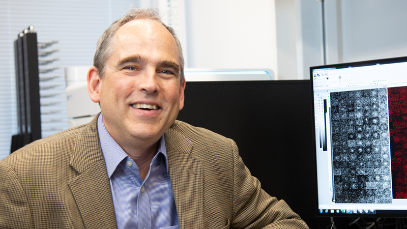
tCycIF, tissue-based cyclic immunofluorescence, is a method for constructing sub-cellular resolution, highly multiplexed images of tumors and tissue. Each “channel” in such an image represents a different molecular marker, and we can routinely perform 40- to 60-channel imaging. This is accomplished by doing the same simple thing over and over (four-color imaging) and building the complete image like a big layer cake—it’s simple when each layer comes out of the oven but becomes really valuable once you assemble it and put on the frosting. We perform tCyCIF using existing instruments and reagents, making it easy for others to implement the approach.
You’re a big proponent of curiosity-driven research. How do you integrate it into your lab?
Breakthroughs often come from orthogonal thinking and unexpected directions. You’ll have someone who isn’t deeply familiar with a field taking a fresh look at a problem and finding something curious about the outcome of an experiment or a clinical trial. If the results of a trial for a particular drug combination treating melanoma, for example, has a success rate of 60%, a curious observer inevitably asks: what about the other 40%? Last December a postdoc in my group, Adam Palmer, and I published a paper in Cell reanalyzing data from a large number of clinical trials of combination therapies. Adam’s background is antibiotic resistance not cancer therapy, and he was curious about the widespread observation that combinations of anti-cancer drugs (like antibiotic combinations) typically work better than single drugs. He showed that overall benefit likely arose from sub-sets of patients who responded to different single drugs in the combination rather than the two drugs at the same time. This model of “independent action” is very simple but has been largely ignored since it was developed 70 years ago.
Based on Adam’s findings, it might often be appropriate to start with a drug mixture, since it is hard to predict response in a particular patient, and then assay an on-treatment biomarker so that only the effective drug is continued. This would reduce side effects and improve outcomes. It was curiosity-driven research by an outsider, not a systematic program of study by an expert, that provided this fresh perspective.
Can you tell us about the Ludwig Cancer Atlas project?
No activity is more important in the routine diagnosis of cancer than the acquisition of biopsies and their examination by pathologists. But the methods currently in use are remarkably old fashioned—it is only in the last year that the FDA has provided guidance on using computer screens rather than microscopes themselves to examine histopathological specimens. The Tumor Atlas project will develop and deploy new approaches to histopathology that promise to revolutionize our understanding of basic cancer biology by providing highly detailed information on the molecular states of tumor, stromal and immune cells. These research applications will drive innovations in diagnostic pathology, which we expect to fully encompass and greatly extend current genomic approaches.
The first phase of the project is to create high-dimensional images of whole tumors so we can precisely locate tumor cells, supporting stroma and immune cells and determine where they interact functionally. Tumor cells will be assayed at a molecular level so we can determine the activity of cancer-causing pathways and look at possible resistance mechanisms. The second phase is taking the resulting picture data and combining the expertise of human pathologists with artificial intelligence algorithms to work out which patients will respond to a therapy or whether their cancer will progress. Right now, we are looking at dozens of tumors of six types, but we envision that in the near future the Cancer Atlas will consist of many more samples so that deep statistical analysis is possible. We will place the Ludwig Cancer Atlas in the public domain so a broad community of scientists and physicians can participate in its interpretation.
How does tCycIF fit into the Cancer Atlas project?
For the first two to three years we expect t-CyCIF to be the dominant method of data collection for the Cancer Atlas. During that time, we will be evaluating possible alternative technologies that might be even more informative. We greatly welcome input from the Ludwig community with respect to alternative or complementary technologies. We also intend to add single cell genomic data to the Atlas, and this will also require new computational and experimental approaches.
What Ludwig sites will be involved in the project?
We expect to engage the broad Ludwig community in the analysis of Atlas data and in making suggestions for future experiments. All of the tissues that we’ve processed so far have come from US hospitals, but we aim to expand this to international sites. For example, once approvals are in place, we aim to study ovarian cancer samples from George Coukos’ lab in Lausanne and Barrett’s esophagus samples from Xin Lu in Oxford. The Ludwig Chicago center will be involved in our training programs. Implementing Atlas technology is well within the expertise of all Ludwig Centers and I suspect that some will want to perform the work locally and others will want to work with the Harvard Center.
How do you envision the collaboration working?
The first step is to get the workflow and analytical software working in Lausanne. The second is to process samples from Lausanne at Harvard while assessing the feasibility of performing tCyCIF in Lausanne. The third is developing panels of antibodies and analytical tools needed to get scientific insights from complex images. Ideally, we’d like to have a joint clinical fellow or an advanced postdoc who would travel back and forth between Lausanne and Harvard on a semi-regular basis. We will be evaluating this working arrangement at six- and 12-month intervals to determine if that is how we’ll also proceed with Xin Lu’s lab. We all intend to meet up in Lausanne this winter to evaluate progress.
What disciplines will be involved in the effort?
There are basically four work streams that we’ll be coordinating. One involves pathologists who guide antibody selection and interpret image data—at the Ludwig Harvard Center we have eight practicing pathologists involved. Second, and equally important, are cell biologists like Joan Brugge with deep knowledge and understanding of the underlying molecular pathways. The third set of activities involves the computer science and artificial intelligence software needed to construct and visualize a “Google Map” of human tumors and tissues. Finally, a team of analytical chemists and automation experts will ensure that we generate consistent and reliable data.
How will it benefit the research community and ultimately the cancer patient?
I think there are two research communities that are going to be impacted. The first is where Ludwig is really strong—translational cancer biology. And the second is the clinical trialists because we’ll be able to take a deeper molecular look at the specimens currently collected as part of many clinical trials. For patients, it’s the promise of getting the right drug to the right individuals. Our best guess is that even at the most advanced medical centers in the world only a third of patients really benefit from the therapy they receive. In some cases, better drugs are not yet available but in many others the challenge is matching patients to the optimal therapy. It is here that advanced histopathology could be very helpful.
Looking out eight or 10 years, we expect that multiple analytical technologies will come together in the design of better clinical trials. Our goal is to improve clinical trial design and interpretation as a way of accelerating drug development and bringing new drugs to market. This will provide new options for patients but it also promises to bring down the cost of developing new medicines, something we are exploring with the FDA and the pharmaceutical industry.
How can interested Ludwig community members become a part of this initiative?
Ludwig members can collaborate with us using Harvard instruments or be trained here so that they can implement tCyCIF in their own labs. They can also learn it on their own from published papers and protocols. All they’ll need is a microscope, antibodies and a few simple chemicals. A key component of this initiative is to make the data and the code freely available to the Ludwig community to demystify and democratize high dimensional histology.
What would you like to accomplish in the next 5 to 10 years?
Cancer, like human physiology in general, is a complex process involving many interacting molecular processes. In general, we do not yet know how to derive actionable information from such complexity when diagnosing and treating cancer. We therefore fall back on simple rules of thumb, such as treating patients continuously at the maximum tolerated dose. My goal is to build more predictive and data driven computational models that manage this complexity and both explain and predict how cells and patients respond to therapy. These are human-in-the loop systems, in which we empower scientists and physicians to exploit a much broader range of data on biological function.
What do you like most about being a scientist?
Ten years ago, I would have told you that it was the freedom to pursue my own ideas and the chance for reinvention. Now, it’s the interaction with students and fellows that is the single most enjoyable aspect of being a scientist. Many of them have very interesting ideas and it’s my role as mentor to help them refine these ideas and test them. I also greatly enjoy helping students and postdocs take ideas from our lab into the commercial world through start-up enterprises.
If you could be a superhero, what would you want your superpowers to be?
Flight, so I could avoid the interminable check-in lines. Time control, to avoid missing deadlines. Telepathy, to understand what reviewer number 3 is complaining about.
If you could have dinner with anyone from history, who would it be and why?
Alan Turing. His ideas about computation and artificial intelligence developed in the 1930s and 1940s are now finding practical implementation. It would be fascinating to get his views on the performance of a Turing Test using contemporary and future technology.
